
Dr.Mohamed Abdel Aal Hussein
- Consultant of pediatric orthopedic surgery and limb reconstruction
- Chair of pediatric orthopedic committee - SICOT

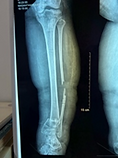
Fibular hemimelia
The most prevalent long bone deficiency is fibular hemimelia.
This condition manifests in a spectrum of severity, ranging from mild fibular deficiency accompanied by limb length inequality (LLI) to significantly short limbs with foot and ankle deformities.
The treatment of fibular hemimelia (without resorting to amputation) seeks to restore a functional limb with nearly equal length to the normal limb at maturity, accompanied by a plantigrade and stable foot. Patients with fibular hemimelia necessitate multiple sessions of limb lengthening. Therefore, surgeons must prioritize understanding the family’s preferences while setting realistic expectations.
Ultimately, patient recommendations must remain individualized, involving extensive discussions of the advantages and disadvantages of available treatments.


Tibial Hemimelia
Tibial hemimelia is a spectrum of deformities characterised by a shortened or absent tibia and a relatively unaffected fibula.
Tibial hemimelia is usually associated with lower extremity abnormalities and other organ system anomalies, most commonly affecting the foot.
The recommended treatment varies according to the type. The vast majority of patients with tibial hemimelia will require surgical treatment of some form for their condition.
Families rarely accept amputation in our culture. There are reconstructive limb salvage procedures available, and in the hands of a skilled surgeon, they can provide favourable outcomes.


Acute osteomyelitis
Acute osteomyelitis can be a devastating or even fatal disease with a high rate of sequelae, especially in resource-poor countries. AHOM is typically a monomicrobial disease, Staphylococcus aureus (S. aureus) remains the most common pathogen globally notwithstanding that Methicillin-resistant Staphylococcus aureus (MRSA) has an increased.
Early diagnosis is the key to successful management and the prevention of long-term sequelae.
the management for AHOM includes Empiric IV (intravenous) antibiotics based on the most likely causative agents, Source control: specimens for microscopy, culture and sensitivity (MCS).
If there is no response to medication within days, an intervention such as draining an abscess might speed up the healing process, A corticotomy can be performed for intraosseous collections, and drains are routinely placed. Bone drilling is required in patients with subperiosteal or intramedullary abscesses
The limb segment should be protected in the first six weeks to prevent a pathological fracture.


Flat foot
Flat foot is one of the commonest foot deformities in children and young adults, with a wide range of severity and symptoms. It is complex and involves three-dimensional deformities with loss of the longitudinal arch height, hindfoot valgus and forefoot abduction.
Surgical treatment is aimed at relieving a patient’s symptoms by realigning the foot to improve foot kinematics during the gait cycle and reduce the possibility of degenerative changes in the hindfoot and midfoot.
Lateral column lengthening remains the most interesting option in children, as it allows further growth and does not involve arthrodesis. It has satisfactory clinical and radiological results, with high patient satisfaction and an acceptable level of complications.


Paralytic valgus ankle
Neurological deficit resulting in the lack of motor control often leads to a valgus position of the feet and ankles.
Valgus can result from deformity in the hindfoot, distal tibia, or both, so careful attention must be paid to determine the precise anatomical location of the deformity.
We should address each component separately, as neglecting to treat all contributing locations of deformity will result in an unsuccessful outcome.


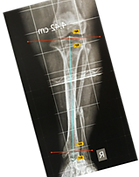
Slipped Capital Femoral Epiphysis (SCFE)
(SCFE) is a frequent cause of a nontraumatic painful hip of the adolescence due to slippage of the metaphysis relative to the epiphysis through the proximal femoral physis.
Slip stabilisation is the main goal of any treatment of SCFE; the choice of surgical management should be made based on acuity, the severity of displacement, stability, and hip morphology.
In situ fixation is the widely accepted treatment with a low incidence of complications.
Recently , Fixation with telescopic screw for SCFE in patients less than 11 years old, with mild to moderate slippage, allows the continuous growth and remodeling of the proximal femur, thus avoiding deformities .

Forearm deformities in Multiple Hereditary Exostoses
Deformations of the forearm in children caused by MHE often progress during their growth period, worsening over time and leading to dysfunction and abnormalities in the elbow and wrist joints.
Ulnar lengthening combined with gradual correction of the radial deformity with open wedge osteotomy effectively restores forearm and wrist alignment, facilitating a satisfactory range of supination, pronation, and active flexion and extension of the wrist.


Blount disease
Blount disease is a multi-planar abnormality consisting of tibia vara, procurvatum, internal rotation, and disordered endochondral ossification of the medial part of the proximal tibial epiphysis; depression of the medial tibial plateau causes joint incongruity and instability.
The goal of treatment in Blount disease is to obtain a well-aligned lower extremity with equal legs and normal joint orientation.
Elevation of the medial tibial plateau with proximal tibial osteotomy is recommended. With this technique, the deformity is corrected, the articular surface is restored, and recurrence can be reduced. mended in severe early-onset Blount disease.


Perthes disease
Perthes disease is an uncommon paediatric disorder characterised by the loss of blood flow to the spherical femoral head of the thigh bone. Consequently, the femoral head undergoes collapse. This procedure may result in the ball becoming flattened. Consequently, Legg-Calvé-Perthes disease may lead to pain and rigidity in the hip joint for a duration.
The treatment is to preserve the spherical shape of the femoral head and avert deformation during the progression of the condition. Treatment choices are contingent upon the severity of hip pain, stiffness, radiographic alterations over time, and the extent of femoral head collapse.
Hip distraction preserves correct head alignment by containing abduction, extension, and neutral rotation. Arthrodiastasis presents a biological paradigm that transcends the mechanical notions of anatomical joint centralisation and mechanical stress protection.
Arthrodiastasis in Perthes disease offers numerous advantages while preserving the normal hip structure.


الدكتور مختص فى
-
حالات الخلع الوليدي لمفصل الفخذ
-
حالات الغياب الخلقي لعظمة الساق
-
حالات الغياب الخلقي لعظمة الشظية
-
حالات الغياب الخلقى الجزئى وقصر عظمة الفخذ
-
حالات الغياب الكلى او الجزئي لعظمة الكعبره بالساعد
-
حالات تسطح القدم
-
حالات القدم المخلبيه او القدم القفداء
-
حالات اعوجاجات وتشوه أعلى عظمة الفخذ
-
حالات انقطاع الدورة الدموية عن رأس عظمة الفخذ
-
حالات زحزحة مركزالنمو لرأس عظمة الفخذ
-
حالات الكسور المضاعفة للأطفال والكبار
-
حالات الالتهابات الصديدية لعظام الأطفال والكبار
-
حالات الكسور سيئة الإلتئام لدى الأطفال والكبار